Zebrafish study highlights contralateral eye’s key role in visual function recovery
GA, UNITED STATES, October 17, 2025 /EINPresswire.com/ -- Optic neuropathies including glaucoma, traumatic injury, ischemic damage, and optic neuritis, are the leading causes of irreversible blindness worldwide; however, effective treatments for optic nerve repair remain elusive despite decades of research . Using zebrafish larvae as a model organism, researchers investigated the cooperative mechanisms between the eyes and the brain that underlie visual function restoration following optic nerve injury. They tracked neural repair in zebrafish larvae via optimized two-photon imaging combined with behavioral tests. The study demonstrates that the contralateral eye supplies critical guidance cues for axonal regrowth and behavioral recovery. These findings highlight the zebrafish as a unique model for uncovering mechanisms of neural circuit repair and offer valuable insights for designing therapeutic strategies against human optic neuropathies.
Traditional mammalian models, including mice, hinder optic nerve regeneration research due to poor regenerative capacity, embryonic opacity, complex anatomy, and labor-intensive tissue processing. By contrast, zebrafish larvae support noninvasive imaging (via transparency) and dynamic in vivo monitoring of inflammation and regeneration—facilitated by conserved glial cells (e.g., microglia) and human-like immune responses involving neutrophils and macrophages. Yet, prior imaging approaches were hindered by pigment interference and low resolution, leaving critical gaps in understanding axonal reconnection and the role of supporting cells in coordinating recovery. To overcome these challenges, researchers aimed to develop a refined platform for visualizing structural and functional regeneration along the entire visual pathway.
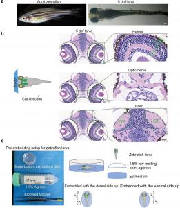
A research team from the Eye Hospital of Wenzhou Medical University has now filled this gap. Their study, published (DOI: 10.1186/s40662-025-00447-z) on August 25, 2025, in Eye and Vision, introduces an advanced in vivo imaging system that combines two-photon microscopy, pigment suppression, and behavioral assays. Using this approach, they demonstrated that the contralateral eye plays a vital role in guiding accurate optic nerve regrowth and restoring visual function after optic nerve injury. The researchers built a longitudinal imaging protocol optimized for zebrafish larvae, employing near-infrared excitation (930 nm), pigment suppression with phenylthiourea, and precise positioning to capture the entire visual pathway. Green fluorescent protein-labeled retinal ganglion cell axons were monitored from retina to tectum, while visual recovery was tested through the optokinetic response (OKR).
Following the removal of the contralateral eye, subsequent induction of optic nerve injury resulted in aberrant optic nerve regrowth and the failure of functional recovery. In contrast, intact contralateral eyes supported organized regrowth and phased restoration of OKR behavior. Quantitative analysis confirmed that recovery in the optic nerve and tectum strongly correlated with improved visual function. Additionally, dual-color imaging revealed the recruitment of neutrophils to the injury site, suggesting immune participation in axonal repair. Together, these experiments provide the first in vivo evidence that contralateral eye cues and immune interactions jointly shape functional optic nerve regeneration, offering new perspectives on how neural circuits reorganize after injury.
“Our findings demonstrate that the contralateral eye is an active contributor to neural repair,” noted Professor Wencan Wu of Wenzhou Medical University. “By visualizing regeneration processes in real time, we confirmed that guidance from the contralateral eye is critical for the accurate reconnection of the optic nerve to the brain. This discovery not only deepens our understanding of regeneration mechanisms in zebrafish but also provides vital insights for developing therapeutic strategies to treat human optic neuropathies and central nervous system injuries—conditions in which spontaneous repair capacity remains highly limited.”
This optimized imaging system establishes zebrafish larvae as a versatile model for dissecting neural repair at both cellular and behavioral levels. By linking axonal regrowth to visual outcomes, the study provides a framework for evaluating candidate regenerative therapies. Future integration with spatial transcriptomics and calcium imaging could reveal the molecular cues driving precise reconnection. These findings may ultimately inform treatments for glaucoma, optic neuritis, and traumatic optic nerve damage. More broadly, the platform highlights evolutionarily conserved repair mechanisms in the central nervous system, offering pathways that could be harnessed to promote functional recovery in humans.
DOI
10.1186/s40662-025-00447-z
Original Source URL
https://doi.org/10.1186/s40662-025-00447-z
Funding Information
This work was supported by the National Key R&D Program of China (Grant No. 2021YFA1101200), the National Natural Science Foundation of China (Grant Nos. 82171048 and 81800842), the Key R&D Program of Wenzhou (Grant No. ZY2022021) and the R&D Program of Wenzhou (Grant No. H20220008), Zhejiang Provincial Natural Science Foundation of China (Grant No. LY22H120005), Research Initiation Funds from Wenzhou Eye Hospital (Grant No. KYQD20221203), and the China Postdoctoral Science Foundation (Grant No. 2023M742674).
Lucy Wang
BioDesign Research
email us here
Legal Disclaimer:
EIN Presswire provides this news content "as is" without warranty of any kind. We do not accept any responsibility or liability for the accuracy, content, images, videos, licenses, completeness, legality, or reliability of the information contained in this article. If you have any complaints or copyright issues related to this article, kindly contact the author above.

